
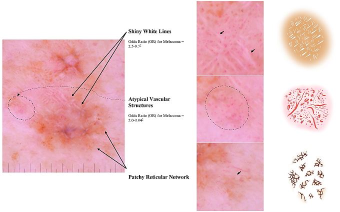
A questionable leg lesion is found in a patient with a history of melanoma. Dotted vessels, shiny white lines, and peripheral tan structureless areas raise concern for melanoma. Which dermoscopic feature has prognostic value?
Diagnosis vs. Prognosis of Melanoma
By Zaeem Nazir, BA, and Ashfaq A. Marghoob, MD
Dermatology Service, Department of Medicine, Memorial Sloan Kettering Cancer Center, New York, New York
CASE HISTORY
A patient in his seventies presents to clinic for periodic skin cancer surveillance examination. He has a personal history of melanoma. He denies any new or changing lesions and describes himself as an “outdoorsy” person. Physical examination reveals a questionable leg lesion. On clinical and dermoscopic exam, you observe the following:
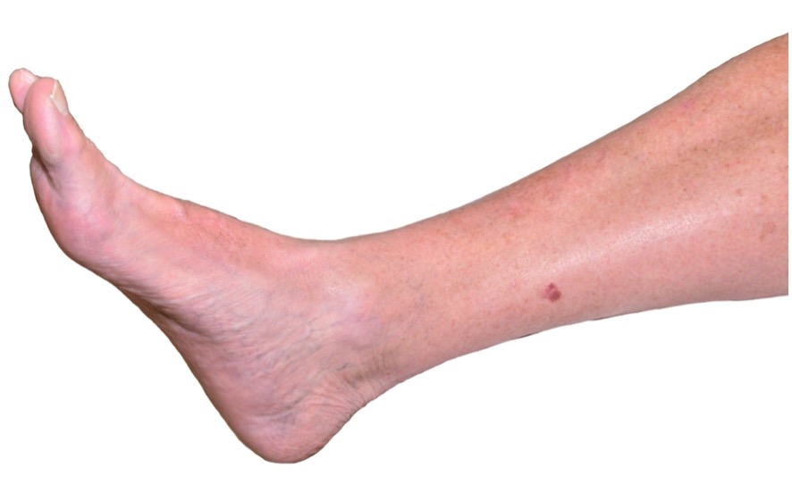
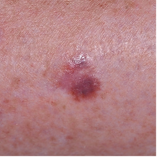
Figures 1a and 1b: Images of clinical lesion
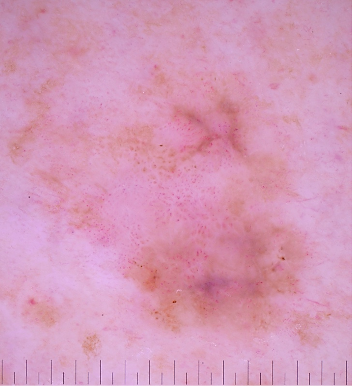
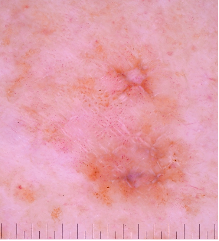
Figures 2a and 2b: Dermatoscopic images of the lesion
Question: Which of the following dermoscopic features provides prognostic information for the lesion in question?
A. Dotted Vessels
B. Shiny White Lines
C. Peripheral Tan Structureless Areas
D. Patchy Reticular Network
E. All of the above
F. None of the above
Answer: B. Shiny White Lines
What Is Your Diagnosis?
DISCUSSION
Although dotted vessels, shiny white lines, and peripheral tan structureless areas raise concern for melanoma,1 shiny white lines are the only dermoscopic feature with potential prognostic value.2 Its presence is associated with increased risk of invasive melanoma relative to in-situ disease.3 Additionally, invasive melanomas with shiny white lines tend to be thicker than those without shiny white lines.2 Shiny white lines are postulated to correspond with the collagen remodeling occurring during the vertical growth phase of melanoma.4,5 Since neoangiogenesis often occurs simultaneously with matrix alteration in melanoma, it is not uncommon to find atypical vessels whenever shiny white lines are present, as seen in this case. This lesion was excised, and pathology confirmed the diagnosis of an invasive melanoma, superficial-spreading type with a thickness of 0.7 mm.
References:
- Marghoob NG, Liopyris K, Jaimes N. Dermoscopy: A Review of the Structures That Facilitate Melanoma Detection. J Osteopath Med. 2019;119(6):380-390. doi:10.7556/jaoa.2019.067
- Verzi AE, Quan VL, Walton KE, et al. The diagnostic value and histologic correlate of distinct patterns of shiny white streaks for the diagnosis of melanoma: A retrospective, case-control study. JAAD. 2018;78(5):913-919. doi:10.1016/j.jaad.2017.11.021
- Balagula Y, Braun RP, Rabinovitz HS, et al. The significance of crystalline/chrysalis structures in the diagnosis of melanocytic and nonmelanocytic lesions. JAAD. 2012;67(2):194.e1-194.e8. doi:10.1016/j.jaad.2011.04.039
- Kääriäinen E, Nummela P, Soikkeli J, et al. Switch to an invasive growth phase in melanoma is associated with tenascin-C, fibronectin, and procollagen-I forming specific channel structures for invasion: TN-C and FN in melanoma dissemination. J Pathol. 2006;210(2):181-191. doi:10.1002/path.2045
- Krenacs T, Kiszner G, Stelkovics E, et al. Collagen XVII is expressed in malignant but not in benign melanocytic tumors and it can mediate antibody induced melanoma apoptosis. Histochem Cell Biol. 2012;138(4):653-667. doi:10.1007/s00418-012-0981-9
Disclosure: This work was supported in part by Memorial Sloan Kettering Cancer Center’s NIH/National Cancer Institute Cancer Center support grant P30 CA008748.