
What started as a roundish macular skin lesion became a painless eschar in a febrile 15-year-old cancer patient who was immunocompromised and immunosuppressed. What is your diagnosis?
Ulcerated Skin Lesion Covered by Eschar in Young Cancer Patient
By Ted Rosen, MD
Professor of Dermatology, Baylor College of Medicine, Houston, Texas
CASE HISTORY
A 15-year-old male patient with known B-cell acute lymphoblastic leukemia (B-ALL) was seen in the emergency department with the chief complaint of a skin lesion on the right arm.
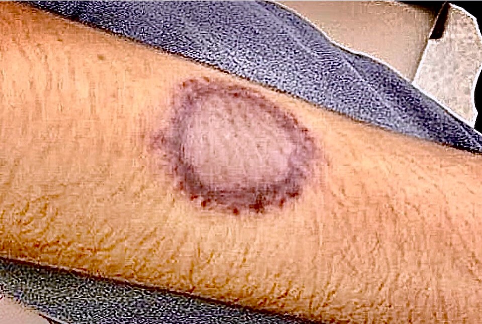
At the time of this visit he was receiving periodic intravenous chemotherapy consisting of vincristine and daunorubicin. At presentation he had a low-grade fever and a round, macular skin lesion. The latter consisted of a slightly dusky center and a peripheral non-blanching violaceous rim (Figure 1).
The emergency department made a diagnosis of “fixed drug eruption” and asked that all medications be temporarily withheld and that triamcinolone be applied to the affected area twice daily.
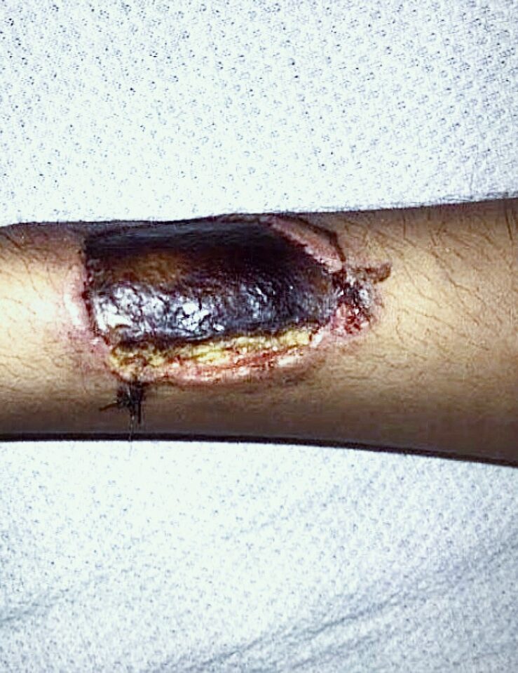
Unfortunately, he returned to the hospital a week later with a remarkably painless enlarged cutaneous lesion and a temperature of 102.6°F and a blood-tinged nasal discharge.
The skin lesion was now notably wider and deeper, ulcerated, and was covered by a prominent eschar (Figure 2).
The patient was started on broad spectrum antibiotics. A biopsy demonstrated a pauci-cellular necrotizing vasculitis.
What is your diagnosis?
DISCUSSION
Due to the emergence of a painless eschar in a febrile individual who was both immunocompromised (aggressive leukemia) and immunosuppressed (multiagent chemotherapy), the presumptive diagnosis of ecthyma gangrenosum was made.
This entity is usually a sign of serious blood-borne infection in an altered host. While the majority (~75%) of cases of ecthyma gangrenosum are due to Pseudomonas septicemia, it is well documented to be the result of a variety of bacterial, fungal, and viral pathogens.
In this case, radiographs of the sinuses showed fungal balls, and culture grew the zygomycete Rhizopus. A culture from the arm lesion also grew Rhizopus.
Treatment was changed to intravenous amphotericin-B.
Recommended Reading:
Ruiz-Sanchez D, Valtueña J, Garabito Solovera E, et al. Ecthyma gangrenosum, beyond Pseudomonas aeruginosa. Enferm Infecc Microbiol Clin (Engl Ed). 2021;39(10):526-527. doi:10.1016/j.eimce.2021.09.005.